

Vol. 14. Núm. 1.
Páginas 11-20 (Enero 2024)

Actualización
Esofagitis infecciosa y esofagitis por cáusticos

Infectious esophagitis and corrosive esophagitis

C. Trueba ColladoA. Castellano LópezM.I. Higuera Fernández
Doi : https://10.1016/j.med.2024.01.002
Opciones
Resumen

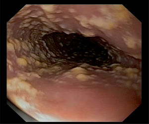
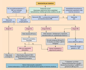
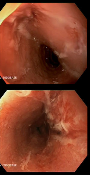
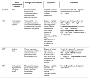
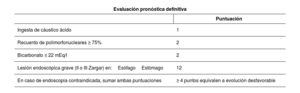
La esofagitis se define como la afectación del revestimiento esofágico caracterizada por inflamación y daño tisular. La esofagitis infecciosa constituye la tercera causa de esta patología a nivel mundial, siendo la esofagitis por Candida la causa más frecuente. Suele aparecer en pacientes inmunodeprimidos, y el diagnóstico se realiza principalmente mediante técnicas endoscópicas y muestras histológicas. La esofagitis por cáusticos es una causa de esofagitis más infrecuente, aunque puede presentarse como una afectación esofágica grave con complicaciones a corto y largo plazo, con gran impacto en la calidad de vida del paciente. La endoscopia tiene un papel relevante en el diagnóstico y el pronóstico con la clasificación de Zargar, tanto en el manejo agudo como en el tratamiento de las complicaciones posteriores.

Palabras clave
Esofagitis infecciosa
Esofagitis cáustica
Disfagia
Abstract

Esophagitis is defined as involvement of the esophageal lining characterized by inflammation and tissue damage. Infectious esophagitis is the third leading cause of this disease worldwide, with esophagitis due to Candida being the most frequent cause. It usually appears in immunosuppressed patients. The diagnosis is made mainly through endoscopic techniques and histological specimens. Corrosive esophagitis is a rarer cause of esophagitis, although it can present as severe esophageal involvement with short- and long-term complications and a big impact on the patient's quality of life. Endoscopy with the Zargar classification plays a relevant role in the diagnosis and prognosis in both acute management and the treatment of subsequent complications.

Keywords
Infectious esophagitis
Corrosive esophagitis
Dysphagia

Identifíquese

¿Aún no es suscriptor de la revista?
Comprar el acceso al artículo

Comprando el artículo el pdf del mismo podrá ser descargado

Teléfono para incidencias

De lunes a viernes de 9h a 18h (GMT+1) excepto los meses de julio y agosto que será de 9 a 15h
Llamadas desde España 932 415 960
Llamadas desde fuera de España +34 932 415 960
Bibliografía
[1]
A.A. Mohamed
202401250544492078
202401250544492079
, X. Lu, F.A. Mounmin.
Diagnosis and treatment of esophageal candidiasis: Current updates.
Can J Gastroenterol Hepatol., (2019), 2019 pp. 3585136
[2]
P. Hoversten
2024012505444920710
2024012505444920711
, A.K. Kamboj, D.A. Katzka.
Infections of the esophagus: An update on risk factors, diagnosis, and management.
Dis Esophagus., (2018), 31
[3]
Khan
2024012505444920712
AN. Infectious (candida, herpes, cytomegalovirus, HIV, tuberculosis) esophagitis imaging [Internet]. 2022 [Consultado 17 Ago 2023]. Disponible en: https://emedicine.medscape.com/article/376127-print.
[4]
L.I. Chen
2024012505444920713
, J.M. Chang, M.C. Kuo, S.J. Hwang, H.C. Chen.
Combined herpes viral and Candidal esophagitis in a CAPD patient: case report and review of literature.
Am J Med Sci., (2007), 333 pp. 191-193
[5]
V. Neri
2024012505444920714
, M. Ahmed, D. Kingsley-Godwin, M.J. Kingsley-Godwin, J. Godwin.
Infectious esophagitis.
En: Esophagitis and gastritis: Recent updates, Intech Open, (2021), pp. 1-18
[6]
P. Hoversten
2024012505444920715
2024012505444920716
, F. Otaki, D.A. Katzka.
Course of esophageal candidiasis and outcomes of patients at a single center.
Clin Gastroenterol Hepatol., (2019), 17
[7]
Y. Takahashi
2024012505444920717
2024012505444920718
, N. Nagata, T. Shimbo, T. Nishijima, K. Watanabe, T. Aoki, et al.
N. Long-term trends in esophageal candidiasis prevalence and associated risk factors with or without HIV infection: Lessons from an endoscopic study of 80,219 patients.
PLoS One., (2015), 10 pp. e0133589
[8]
M. Feldman
2024012505444920719
2024012505444920720
, L.S. Friedman, L.J. Brandt, D.A. Katzka.
Capítulo 45. Trastornos esofágicos causados por fármacos, traumatismos e infecciones.
En: Sleisenger y Fordtran, Enfermedades Digestivas y Hepáticas: Fisiopatología, Diagnóstico, Tratamiento, Elsevier, (2022), pp. 661-669
[9]
C.M. Wilcox
2024012505444920721
.
Esophageal disease in the acquired immunodeficiency syndrome: Etiology, diagnosis, and management.
Am J Med., (1992), 92 pp. 412-421
[10]
P.G. Pappas
2024012505444920722
2024012505444920723
, C.A. Kauffman, D.R. Andes, C.J. Clancy, K.A. Marr, L. Ostrosky Zeichner, et al.
Clinical practice guideline for the management of candidiasis: 2016 update by the Infectious Diseases Society of America.
Clin Infect Dis., (2016), 62 pp. 1-50
[11]
N.K. Ahuja
2024012505444920724
2024012505444920725
, J.O. Clarke.
Evaluation and management of infectious esophagitis in immunocompromised and immunocompetent individuals.
Curr Treat Options Gastroenterol., (2016), 14 pp. 28-38
[12]
J. Ramanathan
2024012505444920726
, M. Rammouni, J. Baran Jr., R. Khatib.
Herpes simplex virus esophagitis in the immunocompetent host: an overview.
Am J Gastroenterol., (2000), 95 pp. 2171-2176
[13]
M. Sánchez Cazalilla
2024012505444920727
, A.J. Lucendo.
Otras esofagitis. Etiología infecciosa y tóxica/cáustica. ESOFAGITIS eosinofílica.
[14]
K. Yagain
2024012505444920728
, L. Rao, K. Pai, G. Pai.
Cytomegalovirus esophagitis in nonimmunocompromised patient presenting as an acute necrotic (black) esophagitis.
J Pathol Microbiol., (2011), 54 pp. 852-853
[15]
M. Rosołowski
2024012505444920729
, M. Kierzkiewicz.
Etiology, diagnosis and treatment of infectious esophagitis.
Prz Gastroenterol., (2013), 8 pp. 333-337
[16]
Robertson
2024012505444920730
K, Nagra N, Mehta D. Esophageal candidiasis - statpearls - NCBI bookshelf [Internet]. 2023 [Consultado 6 Ago 2023]. Disponible en: https://www.ncbi.nlm.nih.gov/books/NBK537268/.
[17]
G. Gençdal
2024012505444920731
, S. Degirmencioglu, M. Akyıldız.
Diffuse esophageal squamous papillomatosis covering the entire esophagus.
Clin Gastroenterol Hepatol., (2018), 16 pp. A28
[18]
J.L. Petrick
2024012505444920732
, A.B. Wyss, A.M. Butler, C. Cummings, X. Sun, C. Poole, et al.
Prevalence of human papillomavirus among oesophageal squamous cell carcinoma cases: Systematic review and meta-analysis.
Br J Cancer., (2014), 110 pp. 2369-2377
[19]
S. Jain
2024012505444920733
, N. Kumar, D.K. Das, S.K. Jain.
Esophageal tuberculosis. Endoscopic cytology as a diagnostic tool.
Acta Cytol., (1999), 43 pp. 1085-1090
[20]
M. Sharma
2024012505444920734
, R.S. Ecka, A. Somasundaram, A. Shoukat, V. Kirnake.
Endoscopic ultrasound in mediastinal tuberculosis.
Lung India., (2016), 33 pp. 129-134
[21]
P.D. Smith
2024012505444920735
, M.S. Eisner, J.F. Manischewitz, V.J. Gill, H. Masur, C.F. Fox.
Esophageal disease in AIDS is associated with pathologic processes rather than mucosal human immunodeficiency virus type 1.
J Infect Dis., (1993), 167 pp. 547-552
[22]
C.M. Wilcox
2024012505444920736
, S.R. Zaki, L.M. Coffield, P.W. Greer, D.A. Schwartz.
Evaluation of idiopathic esophageal ulceration for human immunodeficiency virus.
Mod Pathol., (1995), 8 pp. 568-572
[23]
Hicks
2024012505444920737
C, Clement M. Syphilis: Epidemiology, pathophysiology, and clinical manifestations in patients without HIV [Internet]. 2022 [Consultado 21 Ago 2023]. Disponible en: https://www.uptodate.com/contents/syphilis-epidemiology-pathophysiology-and-clinical-manifestations-in-patients-without-hiv.
[24]
M. Corti
2024012505444920738
, M.F. Villafañe.
El compromiso del esófago en la enfermedad HIV/SIDA.
Acta Gastroent Lat., (2003), 33 pp. 211-220
[25]
J.B. Marshall
2024012505444920739
, R. Singh, T.L. Demmy, J.T. Bickel, E.D. Everett.
Mediastinal histoplasmosis presenting with esophageal involvement and dysphagia: case study.
Dysphagia., (1995), 10 pp. 53-58
[26]
R. Pierre Álvarez
2024012505444920740
, S. Neri, M. Contreras, R. Vásquez, L.C. Ramírez, J.P. Riveros, et al.
Guía de práctica clínica ibero-latinoamericana sobre la esofagitis cáustica en pediatría: Fisiopatología y diagnóstico clínico-endoscópico (1a parte).
Revista Chilena de Pediatría., (2020), 91 pp. 149
[27]
C. Arévalo-Silva
2024012505444920741
, R. Eliashar, J. Wohlgelernter, J. Elidan, M. Gross.
Ingestion of caustic substances: a 15-year experience.
Laryngoscope., (2006), 116 pp. 1422-1426
[28]
M.A.A. De Lusong
2024012505444920742
2024012505444920743
, A.B.G. Timbol, D.J.S. Tuazon.
Management of esophageal caustic injury.
World J Gastrointest Pharmacol Ther., (2017), 8 pp. 90-98
[29]
Derrick Lung
2024012505444920744
M. Caustic ingestions treatment & management [Internet]. Medscape. 2022 [Consultado 15 Ago 2023]. Disponible en: https://emedicine.medscape.com/article/813772-treatment?form=fpf.
[30]
Triadafilopoulos
2024012505444920745
G. Caustic esophageal injury in adults [Internet]. 2022 [Consultado 20 Ago 2023]. Disponible en: https://www-uptodate-com.m-hupa.a17.csinet.es/contents/caustic-esophageal-injury-in-adults?search=causticos&source=search_result&selectedTitle=1∼84&usage_type=default&display_rank=1.
[31]
J.W. Poley
2024012505444920746
, E.W. Steyerberg, E.J. Kuipers, J. Dees, R. Hartmans, H.W. Tilanus, et al.
Ingestion of acid and alkaline agents: outcome and prognostic value of early upper endoscopy.
Gastrointest Endosc., (2004), 60 pp. 372-377
[32]
M. Chirica
2024012505444920747
2024012505444920748
, M.D. Kelly, S. Siboni, A. Aiolfi, C.G. Riva, E. Asti, et al.
Esophageal emergencies: WSES guidelines.
World J Emerg Surg., (2019), 14 pp. 26
[33]
S. Contini
2024012505444920749
, C. Scarpignato.
Caustic injury of the upper gastrointestinal tract: a comprehensive review.
World J Gastroenterol., (2013), 19 pp. 3918-3930
[34]
A. Agarwal
2024012505444920750
, D.N. Srivastava, K.S. Madhusudhan.
Corrosive injury of the upper gastrointestinal tract: The evolving role of a radiologist.
Br J Radiol., (2020), 93 pp. 20200528
[35]
Z. Schmitz
2024012505444920751
, R. Hoffman.
Response to emergency computed tomography evaluation of caustic ingestion.
Dis Esophagus., (2022), 35 pp. doac057
[36]
H.H. Ryu
2024012505444920752
, K.W. Jeung, B.K. Lee, J.H. Uhm, Y.H. Park, M.H. Shin, et al.
Caustic injury: Can CT grading system enable prediction of esophageal stricture?.
Clin Toxicol (Phila)., (2010), 48 pp. 137-142
[37]
S.A. Zargar
2024012505444920753
, R. Kochhar, S. Mehta, S.K. Mehta.
The role of fiberoptic endoscopy in the management of corrosive ingestion and modified endoscopic classification of burns.
Gastrointest Endosc., (1991), 37 pp. 165-169
[38]
K.S. Park
2024012505444920754
.
Evaluation and management of caustic injuries from ingestion of acid or alkaline substances.
Clinical Endoscopy., (2014), 47 pp. 301-307
[39]
ASGE Standards
2024012505444920755
of Practice Committee, J.A. Evans, D.S. Early, N. Fukami, T. Ben-Menachem, V. Chandrasekhara, K.V. Chathadi, et al.
The role of endoscopy in Barrett's esophagus and other premalignant conditions of the esophagus. Standards of Practice Committee of the American Society for Gastrointestinal Endoscopy.
Gastrointest Endosc., (2012), 76 pp. 1087
[40]
M. Chirica
2024012505444920756
, L. Bonavina, M.D. Kelly, E. Safarti, P. Cattan.
Caustic ingestion.
Lancet., (2017), 389 pp. 2041-2052