


Vol. 12. Núm. 36.
Páginas 2153-2162 (Junio 2017)

Actualización
Angina estable

Stable angina

B. Santos González
Doi : https://10.1016/j.med.2017.06.011
Opciones
Resumen
Definición y manifestaciones clínicas

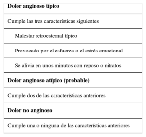
La angina crónica estable es una manifestación común de la enfermedad coronaria, siendo esta última la principal causa de muerte en el mundo. La presencia de angina crónica estable aumenta el riesgo de eventos cardiovasculares mayores, de ahí la importancia de su detección y tratamiento. La angina es tradicionalmente definida como un dolor retroesternal de menos de diez minutos de duración, desencadenado por el ejercicio o situaciones de estrés que cede con el reposo o con nitroglicerina.

Diagnóstico y evaluación

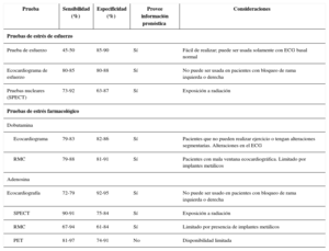
Es importante realizar un diagnóstico correcto, donde es clave la historia clínica; sin embargo, se dispone de un gran número de técnicas diagnósticas tanto invasivas como no invasivas para su correcta detección, que permiten la demostración de isquemia o la visualización de la anatomía coronaria.

Estratificación del riesgo

Tan importante es la realización de un diagnóstico correcto, como el efectuar una adecuada estratificación del riesgo, ya que este proceso sirve para proporcionar una información adecuada sobre el pronóstico y para planificar la actitud terapéutica más adecuada.

Palabras clave
Angina
Enfermedad coronaria
Detección de isquemia miocárdica
Estratificación del riesgo
Abstract
Definition and clinical signs

Chronic stable angina is a common sign of heart disease, which is the primary cause of death worldwide. The presence of chronic stable angina increases the risk of greater cardiovascular events, and thus the importance of their detection and treatment. Angina is traditionally defined as a retroesternal pain lasting for at least 10minutes, triggered by exercise or stressful situations and which recedes when resting or with nitroglycin medication.

Diagnosis and evaluation

It is important to reach an accurate diagnosis, with medical records being crucial. However, there are a great number of diagnostic techniques which are both invasive and non invasive for accurate detection, and which show ischaemia or provide imaging of coronary anatomy.

Risk stratification

It is important to both obtain an accurate diagnosis and complete an appropriate risk stratification, since this process serves to provide the right information about prognosis and to plan the most suitable therapeutic approach.

Keywords
Angina
Coronary heart disease
Detection of myocardial ischaemia
Risk stratification

Identifíquese

¿Aún no es suscriptor de la revista?
Comprar el acceso al artículo

Comprando el artículo el pdf del mismo podrá ser descargado

Teléfono para incidencias

De lunes a viernes de 9h a 18h (GMT+1) excepto los meses de julio y agosto que será de 9 a 15h
Llamadas desde España 932 415 960
Llamadas desde fuera de España +34 932 415 960
Bibliografía
[1]
201706150025508668
D.L. Bhatt, K.A. Eagle, E.M. Ohman, A.T. Hirsch, S. Goto, E.M. Mahoney, et al.
Comparative determinants of 4-year cardiovascular event rates in stable outpatients at risk of or with atherothrombosis.
JAMA., (2010), 304 pp. 1350-1357
[2]
201706150025508669
J. Sanz, P. Moreno, V. Fuster.
The year in atherothrombosis.
J Am Coll Cardiol., (2013), 24 pp. 1131-1143
[3]
2017061500255086610
2017061500255086611
A. Ghattas, H.R. Griffiths, A. Devitt, G.Y. Lip, E. Shantsila.
Monocytes in coronary artery disease and atherosclerosis: where are we now?.
J Am Coll Cardiol., (2013), 62 pp. 1541-1551
[4]
2017061500255086612
2017061500255086613
K. Sakakura, M. Nakano, F. Otsuka, E. Ladich, F.D. Kolodgie, R. Virmani.
Pathophysiology of atherosclerosis plaque progression.
Heart Lung Circ., (2013), 22 pp. 399-411
[5]
2017061500255086614
B. Doyle, N. Caplice.
Plaque neovascularization and antiangiogenic therapy for atherosclerosis.
J Am Coll Cardiol., (2007), 49 pp. 2073-2080
[6]
2017061500255086615
G.A. Lanza, A. Sestito, G.A. Sgueglia, F. Infusino, M. Manolfi, F. Crea, et al.
Current clinical features, diagnostic assessment and prognostic determinants of patients with variant angina.
Int J Cardiol., (2007), 118 pp. 41-47
[7]
2017061500255086616
Y Kitta, J.E. Obata, NakamuraT, M. Hirano, KodamaY, D. Fujioka, et al.
Persistent impairment of endothelial vasomotor function has a negative impact on outcome in patients with coronary artery disease.
J Am Coll Cardiol., (2009), 53 pp. 323-330
[8]
2017061500255086617
W.E. Boden, R.A. O’Rourke, K.K. Teo, P.M. Hartigan, D.J. Maron, W.J. Kostuk, et al.
Optimal medical therapy with or without PCI for stable coronary disease.
N Engl J Med., (2007), 356 pp. 1503-1516
[9]
2017061500255086618
R.M. Califf, P.W. Armstrong, J.R. Carver, R.B. D’Agostino, W.E. Strauss.
27th Bethesda Conference: matching the intensity of risk factor management with the hazard for coronary disease events. Task Force 5. Stratification of patients into high, medium and low risk subgroups for purposes of risk factor management.
J Am Coll Cardiol., (1996), 27 pp. 1007-1019
[10]
G. Montalescot, U. Sechtem, S. Achenbach, F. Andreotti, C. Arden, A. Budaj,
2017061500255086619
2017061500255086620
Task Force Members, et al.
ESC guidelines on the management of stable coronary artery disease: the Task Force on the management of stable coronary artery disease of the European Society of Cardiology.
Eur Heart J., (2013), 34 pp. 2949-3003
[11]
2017061500255086621
G.A. Diamond, J.S. Forrester.
Analysis of probability as an aid in the clinical diagnosis of coronary-artery disease.
N Engl J Med., (1979), 300 pp. 1350-1358
[12]
2017061500255086622
M. Madjid, O. Fatemi.
Components of the complete blood count as risk predictors for coronary heart disease: in-depth review and update.
Tex Heart Inst J., (2013), 40 pp. 17-29
[13]
2017061500255086623
M. Lenzen, L. Ryden, J. Ohrvik, M. Bartnik, K. Malmberg, O.P. Scholte, et al.
Diabetes known or newly detected, but not impaired glucose regulation, has a negative influence on 1-year outcome in patients with coronary artery disease: a report from the Euro Heart Survey on diabetes and the heart.
Eur Heart J., (2006), 27 pp. 2969-2974
[14]
2017061500255086624
A.L. Catapano, I. Graham, G. De Backer, O. Wiklund, M.J. Chapman, H. Drexel, Authors/Task Force Members, et al.
2016 ESC/EAS Guidelines for the Management of Dyslipidaemias.
Eur Heart J., (2016), 37 pp. 2999-3058
[15]
2017061500255086625
J.M. Aroesty, R.G. McKay, G.V. Heller, H.D. Royal, A.V. Als, W. Grossman.
Simultaneous assessment of left ventricular systolic and diastolic dysfunction during pacinginduced ischemia.
Circulation., (1985), 71 pp. 889-900
[16]
2017061500255086626
R.J. Gibbons, J. Abrams, K. Chatterjee, J. Daley, P.C. Deedwania, J.S. Douglas, et al.
ACC/AHA 2002 guideline update for the management of patients with chronic stable angina: summary article: a report of the American College of Cardiology/American Heart Association Task Force on Practice Guidelines (Committee on the Management of Patients With Chronic Stable Angina).
Circulation., (2003), 107 pp. 149-158
[17]
2017061500255086627
R. Gianrossi, R. Detrano, D. Mulvihill, K. Lehman, P. Dubach, A. Colombo, et al.
Exercise-induced ST depression in the diagnosis of coronary artery disease. A meta-analysis.
Circulation., (1989), 80 pp. 87-98
[18]
2017061500255086628
A.P. Morise, G.A. Diamond.
Comparison of the sensitivity and specificity of exercise electrocardiography in biased and unbiased populations of men and women.
Am Heart J., (1995), 130 pp. 741-747
[19]
2017061500255086629
R. Chelliah, B. Anantharam, L. Burden, A. Alhajiri, R. Senior.
Independent and incremental value of stress echocardiography over clinical and stress electrocardiographic parameters for the prediction of hard cardiac events in new-onset suspected angina with no history of coronary artery disease.
Eur J Echocardiogr., (2010), 11 pp. 875-882
[20]
2017061500255086630
2017061500255086631
R. Sicari, P. Nihoyannopoulos, A. Evangelista, J. Kasprzak, P. Lancellotti, D. Poldermans, et al.
Stress echocardiography expert consensus statement: European Association of Echocardiography (EAE) (a registered branch of the ESC).
Eur J Echocardiogr., (2008), 9 pp. 415-437
[21]
2017061500255086632
R. Senior, H. Becher, M. Monaghan, L. Agati, J. Zamorano, J.L. Vanoverschelde, et al.
Contrast echocardiography: evidence-based recommendations by European Association of Echocardiography.
Eur J Echocardiogr., (2009), 10 pp. 194-212
[22]
2017061500255086633
J.U. Voigt, B. Exner, K. Schmiedehausen, C. Huchzermeyer, U. Reulbach, U. Nixdorff, et al.
Strain-rate imaging during dobutamine stress echocardiography provides objective evidence of inducible ischemia.
Circulation., (2003), 107 pp. 2120-2126
[23]
2017061500255086634
L. Imbert, S. Poussier, P.R. Franken, B. Songy, A. Verger, O. Morel, et al.
Compared performance of high-sensitivity cameras dedicated to myocardial perfusion SPECT: a comprehensive analysis of phantom and human images.
J Nucl Med., (2012), 53 pp. 1897-1903
[24]
2017061500255086635
.M. Bateman, G.V. Heller, A.I. McGhie, J.D. Friedman, J.A. Case, J.R. Bryngelson, et al.
Diagnostic accuracy of rest/stress ECG-gated Rb-82 myocardial perfusion PET: comparison with ECG-gated Tc-99m sestamibi SPECT.
J Nucl Cardiol., (2006), 13 pp. 24-33
[25]
2017061500255086636
J.P. Greenwood, N. Maredia, J.F. Younger, J.M. Brown, J. Nixon, C.C. Everett, et al.
Cardiovascular magnetic resonance and single-photon emission computed tomography for diagnosis of coronary heart disease (CE-MARC): a prospective trial.
Lancet., (2012), 379 pp. 453-460
[26]
2017061500255086637
A. Wahl, I. Paetsch, A. Gollesch, S. Roethemeyer, D. Foell, R. Gebker, et al.
Safety and feasibility of high-dose dobutamine-atropine stress cardiovascular magnetic resonance for diagnosis of myocardial ischaemia: experience in 1000 consecutive cases.
Eur Heart J., (2004), 25 pp. 1230-1236
[27]
2017061500255086638
T. Lockie, M. Ishida, D. Perera, A. Chiribiri, K. De Silva, S. Kozerke, et al.
High resolution magnetic resonance myocardial perfusion imaging at 3.0-Tesla to detect hemodynamically significant coronary stenoses as determined by fractional flow reserve.
J Am Coll Cardiol., (2011), 57 pp. 70-75
[28]
2017061500255086639
2017061500255086640
S. Halliburton, A. Arbab-Zadeh, D. Dey, A.J. Einstein, R. Gentry, R.T. George, et al.
Stateof-the-art in CT hardware and scan modes for cardiovascular CT.
J Cardiovasc Comput Tomogr., (2012), 6 pp. 154-163
[29]
2017061500255086641
A.S. Agatston, W.R. Janowitz, F.J. Hildner, N.R. Zusmer, M. Viamonte Jr., R. Detrano.
Quantification of coronary artery calcium using ultrafast computed tomography.
J Am Coll Cardiol., (1990), 15 pp. 827-832
[30]
2017061500255086642
2017061500255086643
I. Danad, Z.A. Fayad, M.J. Willemink, J.K. Min.
New applications of cardiac computed tomography: dual-energy, spectral, and molecular CT imaging.
J Am Coll Cardiol Img., (2015), 8 pp. 710-723
[31]
2017061500255086644
P.K. Vanhoenacker, M.H. Heijenbrok-Kal, R. Van Heste, I. Decramer, L.R. Van Hoe, W. Wijns, et al.
Diagnostic performance of multidetector CT angiography for assessment of coronary artery disease: meta-analysis.
Radiology., (2007), 244 pp. 419-428
[32]
2017061500255086645
J.M. Miller, C.E. Rochitte, M. Dewey, A. Arbab-Zadeh, H. Niinuma, I. Gottlieb, et al.
Diagnostic performance of coronary angiography by 64-row CT.
N Engl J Med., (2008), 359 pp. 2324-2336
[33]
2017061500255086646
2017061500255086647
P.S. Douglas, U. Hoffmann, M.R. Patel, D.B. Mark, H.R. Al-Khalidi, B. Cavanaugh, et al.
Outcomes of anatomical versus functional testing for coronary artery disease.
N Engl J Med., (2015), 372 pp. 1291-1300
[34]
2017061500255086648
J.K. Min, J. Leipsic, M.J. Pencina, D.S. Berman, B.K. Koo, C. Van Mieghem, et al.
Diagnostic accuracy of fractional flow reserve from anatomic CT angiography.
JAMA., (2012), 308 pp. 1237-1245
[35]
S. Windecker, P. Kolh, F. Alfonso, J.P. Collet, J. Cremer, V. Falk,
2017061500255086649
2017061500255086650
Authors/Task Force members, et al.
2014 ESC/EACTS Guidelines on myocardial revascularization: The Task Force on Myocardial Revascularization of the European Society of Cardiology (ESC) and the European Association for Cardio-Thoracic Surgery (EACTS)Developed with the special contribution of the European Association of Percutaneous Cardiovascular Interventions (EAPCI).
Eur Heart J., (2014), 35 pp. 2541-2619
[36]
2017061500255086651
M. Emond, M.B. Mock, K.B. Davis, L.D. Fisher, D.R. Holmes Jr., B.R. Chaitman, et al.
Longterm survival of medically treated patients in the Coronary Artery Surgery Study (CASS) Registry.
Circulation., (1994), 90 pp. 2645-2657
[37]
2017061500255086652
T.D. Miller, V.L. Roger, D.O. Hodge, R.J. Gibbons.
A simple clinical score accurately predicts outcome in a community-based population undergoing stress testing.
Am J Med., (2005), 118 pp. 866-872
[38]
2017061500255086653
D.B. Mark, L. Shaw, F.E. Harrell Jr., M.A. Hlatky, K.L. Lee, J.R. Bengtson, et al.
Prognostic value of a treadmill exercise score in outpatients with suspected coronary artery disease.
N Engl J Med., (1991), 325 pp. 849-853
[39]
2017061500255086654
F.Y. Lin, A.M. Dunning, J. Narula, L.J. Shaw, H. Gransar, D.S. Berman, et al.
Impact of an automated multimodality point-of-order decision support tool on rates of appropriatetesting and clinical decision making for individuals with suspected coronary artery disease: a prospective multicenter study.
J Am Coll Cardiol., (2013), 62 pp. 308-316
[40]
2017061500255086655
K.A. Brown.
Prognostic value of thallium-201 myocardial perfusion imaging. A diagnostic tool comes of age.
Circulation., (1991), 83 pp. 363-381