

Vol. 14. Núm. 8.
Páginas 455-460 (Abril 2024)

Protocolos de práctica asistencial
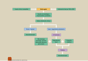
Protocolo diagnóstico de la ileítis aguda

Diagnostic protocol for acute ileitis

V. FloresP. Saralegui
Doi : https://10.1016/j.med.2024.04.017
Opciones
Resumen

La ileítis aguda es una inflamación de la mucosa ileal, habitualmente a nivel del íleon terminal. Es una patología a menudo relacionada con la enfermedad de Crohn. Sin embargo, las causas que pueden dar lugar a la inflamación de la mucosa ileal son múltiples y, a menudo, hacer el diagnóstico diferencial de estas entidades es un reto para el clínico. Dentro de este diagnóstico se debe pensar en enfermedades autoinmunes, fármacos, infecciones, enfermedades por vecindad o neoplasias, entre otras. Diferentes pruebas radiológicas (ecografía abdominal, tomografía computarizada, etc.), endoscópicas, histológicas y de laboratorio (bioquímica o microbiología), además de una correcta anamnesis nos ayudarán a poder orientar a nuestro paciente, evitando la toma de fármacos no indicados o el retraso en el inicio del tratamiento.

Palabras clave
Ileítis aguda
Enfermedad de Crohn
Ileítis infecciosa
Tuberculosis
Abstract

Acute ileitis is an inflammation of the ileal mucosa, usually in the terminal ileum area. It is a disease often related to Crohn's disease. However, there are multiple causes that can give rise to ileal mucosal inflammation and making a differential diagnosis among these entities is often a challenge for clinicians. Within this diagnosis, autoimmune diseases, drugs, infections, environmental diseases, or neoplasms, among others, should be considered. Different radiological (abdominal ultrasound, computed tomography, etc.), endoscopic, histological, and laboratory tests (biochemistry or microbiology) in addition to a proper case history will help clinicians be able to guide patients, avoiding the use of drugs that are not indicated or a delay in starting treatment.

Keywords
Acute ileitis
Crohn's disease
Infectious ileitis
Tuberculosis

Identifíquese

¿Aún no es suscriptor de la revista?
Comprar el acceso al artículo

Comprando el artículo el pdf del mismo podrá ser descargado

Teléfono para incidencias

De lunes a viernes de 9h a 18h (GMT+1) excepto los meses de julio y agosto que será de 9 a 15h
Llamadas desde España 932 415 960
Llamadas desde fuera de España +34 932 415 960
Bibliografía
[1]
M.A. Molina Gutiérrez, A. Vigara, E. Martínez-Ojinaga Nodal, M. Bueno Barriocanal, R. López López.
Value of abdominal ultrasound for evaluation of acute infectious ileitis in children.
An Pediatr (Engl Ed)., (2022), 96 pp. 360-361
[2]
E. Garrido
202405010519423908
202405010519423909
, A.L. Sanromán, M.A. Rodríguez-Gandía, M. Rivero, A. Royuela, V. Moreira, et al.
Optimized protocol for diagnosis of acute ileitis.
Clin Gastroenterol Hepatol., (2009), 7 pp. 1183-1188
[3]
I. Marín-Jiménez, L.A. Menchén, F. Gomollón.
Diagnóstico diferencial de la enfermedad inflamatoria intestinal.
Elsevier, (2010),
[4]
S. Danese, G. Fiorino, J.Y. Mary, P.L. Lakatos, G. D’Haens, L. Moja, et al.
Development of red flags index for early referral of adults with symptoms and signs suggestive of crohn's disease: An IOIBD Initiative.
J Crohns Colitis., (2015), 9 pp. 601-606
[5]
J.B. Puylaert, F.M. Van der Zant, J.A. Mutsaers.
Infectious ileocecitis caused by Yersinia, Campylobacter, and Salmonella: clinical, radiological and US findings.
Eur Radiol., (1997), 7 pp. 3-9
[6]
H. Haskell, C.W. Andrews Jr., S.I. Reddy, K. Dendrinos, F.A. Farraye, A.F. Stucchi, et al.
Pathologic features and clinical significance of “backwash” ileitis in ulcerative colitis.
Am J Surg Pathol., (2005), 29 pp. 1472-1481
[7]
D.T. Patil, R.D. OdzeBackwash.
Is Hogwash: The clinical significance of ileitis in ulcerative colitis.
Am J Gastroenterol., (2017), 112 pp. 1211-1214
[8]
S. DiLauro
2024050105194239010
x4, N.F. Crum-Cianflone.
Ileitis: When it is not Crohn's disease.
Curr Gastroenterol Rep., (2010), 12 pp. 249-258
[9]
B.N. Brewer, D.L. Kamen.
Gastrointestinal and hepatic disease in systemic lupus erythematosus.
Rheum Dis Clin North Am., (2018), 44 pp. 165-175
[10]
M. De Vos, C. Cuvelier, H. Mielants, E. Veys, F. Barbier, A. Elewaut.
Ileocolonoscopy in seronegative spondylarthropathy.
Gastroenterology., (1989), 96 pp. 339-344
[11]
D.Y. Graham, A.R. Opekun, F.F. Willingham, W.A. Qureshi.
Visible small-intestinal mucosal injury in chronic NSAID users.
Clin Gastroenterol Hepatol., (2005), 3 pp. 55-59
[12]
E.V. Marietta, A. Cartee, A. Rishi, J.A. Murray.
Drug-induced enteropathy.
Dig Dis., (2015), 33 pp. 215-220
[13]
S.C.L. Smith, D. Zardo, R. Cannatelli, N. Stevens, M. Iacucci.
Endoscopic findings of checkpoint inhibitor-induced ileitis with use of the latest advanced endoscopic optical diagnosis: near-focus narrow-band imaging.
VideoGIE., (2019), 4 pp. 133-135
[14]
T. Aparicio, A. Zaanan, M. Svrcek, P. Laurent-Puig, N. Carrere, S. Manfredi, et al.
Small bowel adenocarcinoma: epidemiology, risk factors, diagnosis and treatment.
Dig Liver Dis., (2014), 46 pp. 97-104
[15]
G. Mussan-Chelminsky, P. Vidal-González, E. Núñez-García, L.C. Valencia-García, M.A. Márquez-Ugalde.
Intestinal carcinoid tumour: Case report.
Cir Cir., (2015), 83 pp. 438-441
[16]
M.M. Bismar, F.A. Sinicrope.
Radiation enteritis.
Curr Gastroenterol Rep., (2002), 4 pp. 361-365
[17]
P. Chen, L. Anderson, K. Kuixing Zhang, G.A. Weiss.
Eosinophilic gastritis/gastroenteritis.
Current Gastroenterology Reports., (2021), 30;23 pp. 13
[18]
E.C. Ebert, M. Nagar.
Gastrointestinal manifestations of amyloidosis.
Am J Gastroenterol., (2008), 103 pp. 776-787